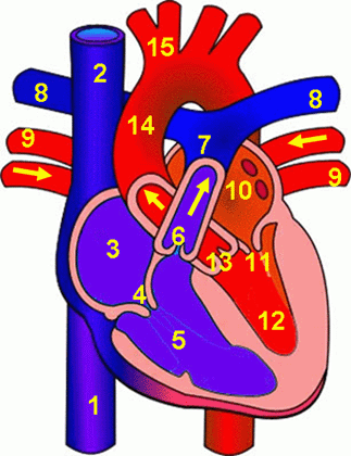
45 structure of the heart without labels
Anatomy of a Human Heart - uofmhealth Located between the lungs in the middle of the chest, the heart pumps blood through the network of arteries and veins known as the cardiovascular system. It pushes blood to the body's organs, tissues and cells. Blood delivers oxygen and nutrients to every cell and removes the carbon dioxide and other waste products made by those cells. Human Heart Diagram Without Labels | Human heart diagram, Heart diagram ... This exhibit depicts the anatomy of the inferior skull including: the foramen magnum, occipital condyles, mastoid process, styloid process, mandibular fossa, palatine bone, sphenoid bone, carotid canal, and the jugular fossa. E Emely cale Pish Posh
Prokaryotic Cells | BioNinja Prokaryotes are organisms whose cells lack a nucleus ('pro' = before ; 'karyon' = nucleus). They belong to the kingdom Monera and have been further classified into two distinct domains: Archaebacteria – found in extreme environments like high temperatures, salt concentrations or pH (i.e. extremophiles); Eubacteria – traditional bacteria including most known pathogenic forms …

Structure of the heart without labels
diagram of heart without labels 13 Best Images of Hip Anatomy Of The Worksheet - Sunflower Anatomy. 11 Pics about 13 Best Images of Hip Anatomy Of The Worksheet - Sunflower Anatomy : u414adad: heart diagram without labels, Congestive Heart Failure: The Essence of Heart Failure Course | CEUfast and also 13 Best Images of Hip Anatomy Of The Worksheet - Sunflower Anatomy. Heart anatomy: Structure, valves, coronary vessels | Kenhub Heart anatomy. The heart has five surfaces: base (posterior), diaphragmatic (inferior), sternocostal (anterior), and left and right pulmonary surfaces. It also has several margins: right, left, superior, and inferior: The right margin is the small section of the right atrium that extends between the superior and inferior vena cava . How Strategy Shapes Structure - Harvard Business Review Summary. Reprint: R0909H. When executives develop corporate strategy, they nearly always begin by analyzing the industry or environmental conditions in which they operate and the strengths and ...
Structure of the heart without labels. Heart Anatomy: Labeled Diagram, Structures, Blood Flow ... - EZmed Chambers of the Heart Let's begin with the chambers of the heart. There are 4 chambers, labeled 1-4 on the diagram below. To help simplify things, we can convert the heart into a square. We will then divide that square into 4 different boxes which will represent the 4 chambers of the heart. draw and label the heart Heart Diagram | Anatomy Of Heart | Different Parts Of The Heart we have 35 Pictures about Heart Diagram | Anatomy Of Heart | Different Parts Of The Heart like 31 Human Heart To Label - Labels Design Ideas 2020, 34 Draw A Heart And Label The Chambers And Valves - Labels For Your Ideas and also how to draw label diagram of heart - Science - Life Processes - 12575. 4 Song Structure Types to Know & When to Use Them in Your … Apr 28, 2022 · This is self-explanatory — the intro is the introduction to the song. And it’s one of the most important parts. According to Music Machinery, about 35% of listeners will skip a song within the first 30 seconds and nearly half of listeners skip a song before it’s over.That’s why your intro has to grab the listener’s ear and hold onto it. SGLT2 inhibitor - Wikipedia SGLT2 inhibitors, also called gliflozins or flozins, are a class of medications that modulate sodium-glucose transport proteins in the nephron (the functional units of the kidney), unlike SGLT1 inhibitors that perform a similar function in the intestinal mucosa.The foremost metabolic effect of this is to inhibit reabsorption of glucose in the kidney and therefore lower blood sugar.
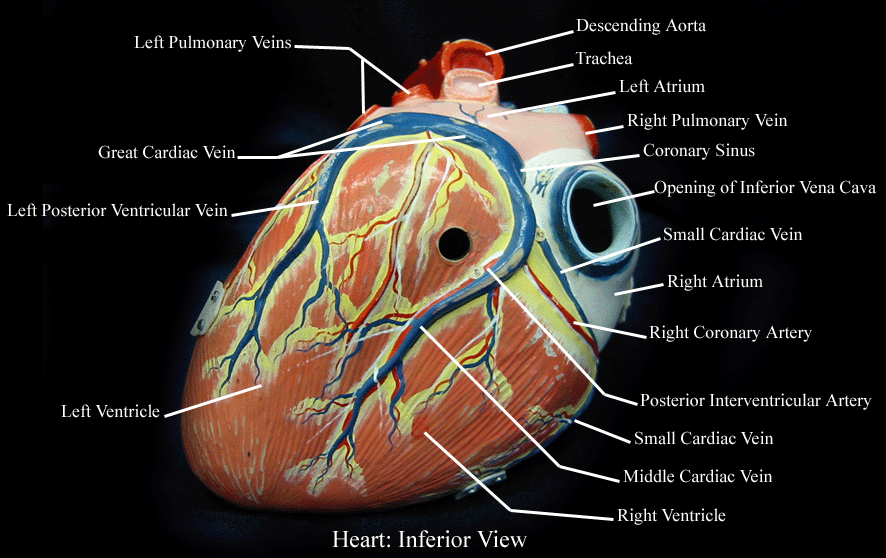
Heart Murmur Detection from Phonocardiogram Recordings: The … Feb 22, 2022 · The segmentation annotation file (with .tsv extension and in plain text format) is composed of three distinct columns: the first column corresponds to the time instant (in seconds) where the wave was detected for the first time, the second column corresponds to the time instant (in seconds) where the wave was detected for the last time, and the third column corresponds … The Anatomy of the Heart, Its Structures, and Functions - ThoughtCo The heart is the organ that helps supply blood and oxygen to all parts of the body. It is divided by a partition (or septum) into two halves. The halves are, in turn, divided into four chambers. The heart is situated within the chest cavity and surrounded by a fluid-filled sac called the pericardium. This amazing muscle produces electrical ... Diagram of the human heart royalty-free images - Shutterstock 14,700 diagram of the human heart stock photos, vectors, and illustrations are available royalty-free. See diagram of the human heart stock video clips. Set goals and get predicted insights based on performance. Heart: Anatomy and Function - Cleveland Clinic The parts of your heart are like the parts of a house. Your heart has: Walls. Chambers (rooms). Valves (doors). Blood vessels (plumbing). Electrical conduction system (electricity). Heart walls Your heart walls are the muscles that contract (squeeze) and relax to send blood throughout your body.
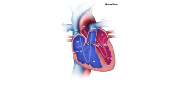
Human Heart (Anatomy): Diagram, Function, Chambers, Location in Body Human Heart (Anatomy): Diagram, Function, Chambers, Location in Body The right atrium receives blood from the veins and pumps it to the right ventricle. The right ventricle receives blood from the... The Heart | Boundless Anatomy and Physiology | | Course Hero The myocardium is the muscle tissue of the heart, composed of cardiac muscle cells called cardiomyocytes that receive nervous stimulation from the sinoatrial (SA) and atrioventricular (AV nodes via the Purkinje fibers. Cardiomyocytes are shorter than skeletal myocytes, and contain fewer nuclei. Cardiac muscle is striated. circulatory system worksheet without labels - Google Search | Heart ... The areas of the heart with MORE oxygen are labeled with an "R". Students will color these areas RED. The areas of the heart with LESS oxygen are labeled with a "B". Students will color these areas BLUE. This diagram is a excellent way to visually represent the direction of blood flow through the heart. The structure of the heart - Structure and function of the heart ... The structure of the heart. If you clench your hand into a fist, this is approximately the same size as your heart. It is located in the middle of the chest and slightly towards the left.

Free Blank Heart Diagram, Download Free Blank Heart Diagram png images, Free ClipArts on Clipart ...
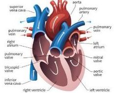
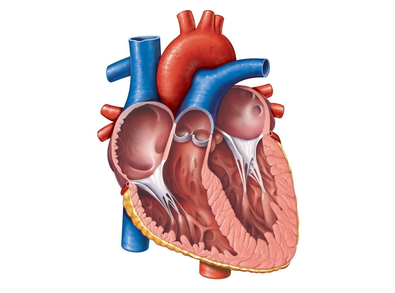
Human Heart Diagram Labeled | Science Trends The heart's atrioventricular valves are structures that join the atria and ventricles of the heart together. This group of valves is comprised of the tricuspid valve and the mitral valve. Beyond this, there is a structure referred to as the aortic valve which separates the left ventricle and the aorta.

Biology A&P Lab: Study guide for quiz on Feb.25th
Heart Diagram with Labels and Detailed Explanation - BYJUS Diagram of Heart. The human heart is the most crucial organ of the human body. It pumps blood from the heart to different parts of the body and back to the heart. The most common heart attack symptoms or warning signs are chest pain, breathlessness, nausea, sweating etc. The diagram of heart is beneficial for Class 10 and 12 and is frequently ...
Get Structure Of Heart Diagram Gcse Background | World of Images
heart diagram without labels heart diagram without labels 13+ heart diagram templates - sample, example, format download. Heart label worksheets diagram human anatomy sparklebox science body ks2 labeling physiology nursing system circulatory diagrams study. Heart diagram label parts template sheet format sample example student templates response blood
Congenital Heart Defects - How the Heart Works | CDC
Cardiovascular System - Heart - Building a Medical Terminology Foundation The walls of the heart consist of three layers: The outer epicardium, which is another name for the visceral pericardium mentioned above. The thick, middle myocardium, which is made of muscle tissue and gives the heart its ability to contract. The inner endocardium, which lines the heart chambers and is the main component of the heart valves.

Human Heart Diagram Without Labels | Human heart diagram, Heart diagram ... This is Page 32 of a photographic atlas I created as a laboratory study resource for my BIOL 121 Anatomy and Physiology I students on the bones and bony landmarks of the axial skeleton. Credits: All photography, text, and labels by Rob Swatski, Assistant Professor of Biology, Harrisburg Area Community College - York Campus, York, PA.
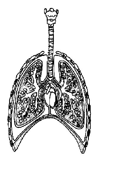
The Anatomy and Physiology of Animals/Respiratory System Worksheet - WikiEducator
Human Heart - Diagram and Anatomy of the Heart - Innerbody The heart is a muscular organ about the size of a closed fist that functions as the body's circulatory pump. It takes in deoxygenated blood through the veins and delivers it to the lungs for oxygenation before pumping it into the various arteries (which provide oxygen and nutrients to body tissues by transporting the blood throughout the body).
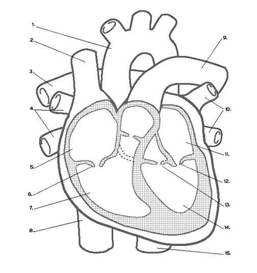
Heart Labeling (Internal)
PDF HEART - STRUCTURE - BiologyMad HEART - STRUCTURE • 4 sections Left atrium Right atrium Left ventricle Right ventricle • heart ry artery Pulmonary vein EAS the blood from he left hand side has to be pumped all around the body. • 2 lo heart Atrioventricular valves - between the atrium and the ventricles Semi-lunar valves - in the pulmonary artery and the aorta
Free Unlabeled Heart Diagram, Download Free Clip Art, Free Clip Art on Clipart Library
A Labeled Diagram of the Human Heart You Really Need to See The human heart, comprises four chambers: right atrium, left atrium, right ventricle and left ventricle. The two upper chambers are called the left and the right atria, and the two lower chambers are known as the left and the right ventricles. The two atria and ventricles are separated from each other by a muscle wall called 'septum'.
12+ Model Heart Labeled | Robhosking Diagram
Label the heart — Science Learning Hub In this interactive, you can label parts of the human heart. Drag and drop the text labels onto the boxes next to the diagram. Selecting or hovering over a box will highlight each area in the diagram. Right ventricle Right atrium Left atrium Pulmonary artery Left ventricle Pulmonary vein Semilunar valve Vena cava Aorta Download Exercise Tweet

CLASS BLOG: BIO 202 Heart Anatomy Worksheet
Losartan - StatPearls - NCBI Bookshelf Feb 09, 2022 · Losartan is FDA approved for the treatment of several medical conditions, which include the following: hypertension, diabetic nephropathy. ARBs are known to be renoprotective in type 2 diabetes mellitus. In hypertension with left ventricular hypertrophy, losartan inhibits angiotensin II-induced cardiac remodeling. It reduces the risk of stroke in these patients. This …

Kenya Forensics Online Resource: CARDIAC MUSCLE TISSUE
Welcome to CK-12 Foundation | CK-12 Foundation FlexBook Platform®, FlexBook®, FlexLet® and FlexCard™ are registered trademarks of CK-12 Foundation.
Know the structure of the heart - Labelled diagram
heart without label Vector Winner Golden Label 458912 Vector Art At Vecteezy . winner vector golden label vecteezy. Netizens Have Said TXT YeonJun Looks Like This Former Wanna One Member . txt yeonjun member wanna netizens former said looks kpopmap enregistrée depuis. Normal Chest Axial Anatomy - Plain And Labeled Sections - YouTube
Label the heart - Teaching resources
heart diagram without labels Label The Heart Worksheets (SB6634) - SparkleBox . heart label worksheets diagram human anatomy sparklebox science body ks2 labeling physiology nursing system circulatory diagrams preschool study. Heart anatomy interior labels section cross blood vessels. Free blank heart diagram, download free blank heart diagram png images.

38 Label The Following Diagram Of The Heart - Labels 2021
Heart Labeling Quiz: How Much You Know About Heart Labeling? Here is a Heart labeling quiz for you. The human heart is a vital organ for every human. The more healthy your heart is, the longer the chances you have of surviving, so you better take care of it. Take the following quiz to know how much you know about your heart. Questions and Answers. 1.
Gorgeous Heart Clipart Diagram - Best Free Clipart : Best Free Clipart
The Anatomy of the Heart - Quiz 1 - Free Anatomy Quiz The circulatory system - lower body image, with blank labels attached. The circulatory system - a PDF file of the upper and lower body for printing out to use off-line. Describe and explain the function of the circulatory system - The circulatory system consists of the heart, the blood vessels (veins, arteries, and capillaries), and the blood.
Post a Comment for "45 structure of the heart without labels"